 At the start of the 1980s, Dr Majid Sheykhzade was a hard-working, academically gifted teenager looking forward to university in Iran. His accolades included a front-page newspaper story that recognized him as a "super-student." However, the devastating Iran-Iraq War caused the closure of all universities, Dr Sheykhzade's options became extreme and lmited: military service or immigration. At the age of 20, Dr Sheykhzade made the difficult choice to leave.
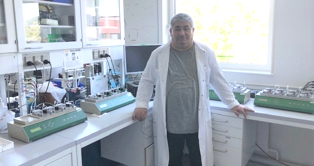
Dr Sheykhzade arrived in Denmark by chance. The majority of his relatives relocated to California, USA. Dr Sheykhzade is grateful that Denmark became his new home: "Denmark is an amazing, unique country. I am pleased to be able to give back to the community that has welcomed me with so much support and opportunity." Links between the cardiovascular system and common diseases Today, as an Associate Professor in the Department of Drug Design and Pharmacology at the University of Copenhagen, Denmark, Dr Sheykhzade is a researcher and educator. His cutting-edge cardiovascular physiology and pharmacology research explores the complex mechanism behind the regulation of arterial tone in the cardiovascular system to understand its role in diseases such as diabetes, stroke, hypertension, congestive heart failure, migraine, and age-related changes. Dr Sheykhzade's research has been helped by a long-standing and fruitful collaboration with a range of researchers that includes Professor Lars Edvinsson, wo is the Director of the Glostrup Research Institute – Rigshospitalet and his team, as well as two of the Institute's key Senior Researchers: Dr Anette Sams and Dr Kristian Agmund Haanes. To date, Dr Sheykhzade has almost 100 papers published in peer-reviewed reputed international scientific journals (such as the British Journal of Pharmacology), as well as several book chapters. As an advocate for the importance of research, he is dedicated to nurturing the next generation of scientific leaders. He also plays a leadership role in the scientific community, providing his time to help advance scientific research and discovery. His community work includes presenter, organizer, and chair of scientific meetings, he is a full member of British Pharmacological Society (BPS), Scandinavian Physiological Society (SPS), and Danish Heart Association. The start of Dr Sheykhzade's career In the 1990s, Dr Sheykhzade's early tertiary education and work experience gave him an understanding and insight into science in the public and private sectors. He obtained a Masters MSc in Pharmacy from the Royal Danish School of Pharmacy, worked at Bispebjerg Hospital Pharmacy, the French pharmaceutical company IPSEN Scandinavia A/S and later in a medical department monitoring clinical trials. In 1996, his growing passion for scientific research led to a 4 year self-financing Ph.D. at the University of Copenhagen. He was able to apply for and attract grants, a skill that he still applies for emerging research. He studied under Prof. Niels Chresten Berg Nyborg, who "introduced me to the fascinating and interesting field of vascular physiology and pharmacology". How DMT Myograph systems fuelled Dr Sheykhzade's research Dr Sheykhzade started using DMT Myograph systems to control and study isolated organ activity and contraction. In addition to using tissue from various rodents, he has also used human subcutaneous resistance arteries. The extensive range of studies includes measuring intracellular calcium concentration using a single chamber myograph system and fluorescence microscopy. Dr Sheykhzade describes the DMT Myographs as fundamental to his research, "DMT myographs are everything to my work. I have succeeded in reaching important milestones in my studies on vascular tone in health and disease by using these robust, reliable organ baths. It happens often that scientific results published in well-reputed journals with high impact factors such as Nature are hardly reproducible by other investigators or laboratories. "My results using myographs are reproducible, it has been proven more than once." "One of the great advantages of the myograph setup is that it can be easily adapted or modified in order to answer many scientific questions. For example, it can be used for electrical field stimulation, placed on the stage of inverted microscope for measurement of intracellular calcium, and used for studying contractions in bladder smooth muscle strips." Experiments that develop critical thinking Dr Sheykhzade introduced the 4-channel wire myograph systems for studying in-vitro techniques in pharmacology to the Master's program. To date, he has supervised approximately 60 Master students as well as 15 PhD students. "I have used myographs in the teaching of both bachelor and Master students with great success. My students love to work with isolated arteries. They enthusiastically add compounds to the myograph baths, record the effects and perform the concentration-response curves. "The myography technique encourages students to be proactive and ask critical questions during experiments. My students become familiar with pitfalls and important tricks when conducting in-vitro experiments with isolated organs. They become skilled at interpreting data, making conclusions, and troubleshooting problems, "said Dr Sheykhzade. Dr Sheykhzade's commitment to teaching and helping new scientists has led to faculty awards and nominations, including two times winner of the best teacher of the year. Future directions in research and education Continuing his scientific endeavors, Dr Sheykhzade currently investigates 1) the role of calcitonin gene-related peptides in the pathophysiology of migraine and 2) the role of formyl peptide receptors (FPRs), toll-like receptors (TLR2 and TLR4) in the development of atherosclerosis. He also looks at vasomotor activity, calcium handling, and metabolism in arteries isolated from different vascular beds of type 2 diabetic mice and rats. Dr Sheykhzade plans to help set up Ph.D. and postdoc projects to conduct investigations on these research projects. "I am working to uncover the complex mechanism behind the inflammatory process that leads to vascular inflammation, atherosclerosis, and ischemia in diabetic patients, especially those with type II diabetes. The key players involved in this inflammatory process are indeed potential targets for drug intervention, and the information obtained from the study will hopefully help early diagnosis and improve the treatment of these patients. "My research at the University of Copenhagen continues to excite me, and DMT myographs are playing an important part. The myographs are cited in 95% of my publications, and there are many more papers ahead. I also want to keep helping young people to find their research passion. We still have many questions to answer." A SELECTION OF DR MAJID SHEYKHZADE'S RECENT PAPERS Pirfenidone Is a Vasodilator: Involvement of KV7 Channels in the Effect on Endothelium-Dependent Vasodilatation in Type-2 Diabetic Mice. Beck L, Pinilla E, Arcanjo DDR, Hernanz R, Prat-Duran J, Petersen AG, Köhler R, Sheykhzade M, Comerma-Steffensen S, Simonsen U. Front Pharmacol. 2021 Jan 12;11:619152. doi: 10.3389/fphar.2020.619152. eCollection 2020. PMID: 33643042 Effect of increased potassium intake on the renin-angiotensin-aldosterone system and subcutaneous resistance arteries: a randomized crossover study. Dreier R, Abdolalizadeh B, Asferg CL, Hölmich LR, Buus NH, Forman JL, Andersen UB, Egfjord M, Sheykhzade M, Jeppesen JL. Nephrol Dial Transplant. 2020 Dec 6:gfaa292. doi: 10.1093/ndt/gfaa292. Online ahead of print. PMID: 33280043 Endothelial Dysfunction and Passive Changes in the Aorta and Coronary Arteries of Diabetic db/db Mice. Beck L, Su J, Comerma-Steffensen S, Pinilla E, Carlsson R, Hernanz R, Sheykhzade M, Danielsen CC, Simonsen U. Front Physiol. 2020 Jun 23;11:667. doi: 10.3389/fphys.2020.00667. eCollection 2020. PMID: 32655412 Fluorescent Analogues of Human α-Calcitonin Gene-Related Peptide with Potent Vasodilator Activity. Zhu J, Pedersen MD, Ahmed LS, Abdolalizadeh B, Grell AS, Berg JO, Thulstrup PW, Franzyk H, Edvinsson L, Sams A, Sheykhzade M, Hansen PR. Int J Mol Sci. 2020 Feb 17;21(4):1343. doi: 10.3390/ijms21041343. PMID: 32079247. Hypermetabolism and impaired endothelium-dependent vasodilation in mesenteric arteries of type 2 diabetes mellitus db/db mice. Arildsen L, Andersen JV, Waagepetersen HS, Nissen JBD, Sheykhzade M. Diab Vasc Dis Res. 2019 Nov;16(6):539-548. doi: 10.1177/1479164119865885. Epub 2019 Jul 31. PMID: 31364402 C-fibers may modulate adjacent Aδ-fibers through axon-axon CGRP signaling at nodes of Ranvier in the trigeminal system. Edvinsson JCA, Warfvinge K, Krause DN, Blixt FW, Sheykhzade M, Edvinsson L, Haanes KA.J Headache Pain. 2019 Nov 12;20(1):105. doi: 10.1186/s10194-019-1055-3.PMID: 31718551 By Helen Inkson | Communications Manager |DMT-APAC   Myography is the go-to methodology of choice for assessing blood vessel function and cardiovascular dysfunction in safety pharmacology. The idea behind this principle is that small or large blood vessel ring preparations are mounted using wires or pins secured unto two supports. The first support is connected to a micrometer to vary the vessel diameter, and the other support is attached to a force transducer. This setup allows the vessel to generate a standardized basal tone on all experimental ring preparations in a process called normalization. For large conduit arteries, this is less applicable.
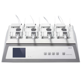
Why is normalization important for all myography studies in small resistance arteries? Normalization imparts a measured optimal passive tension on the vessel before experimentation. This is a pre-condition to generating maximum vessel contraction. To understand this principle, we need to revisit the sliding filament mechanism of actin-myosin cross-bridge in smooth muscle contraction. In brief, thin actin filaments interact optimum with thick myosin filaments to form cross-bridges. At the ideal level, the vessel is not overstretched nor under-stretched. This ideal condition generates maximum vascular contraction. Perfecting and replicating this optimum tension on each vessel for experimentation is the holy grail of vascular function in resistance arteries. This is critical for reliable, consistent, and reproducible results for better data interpretation. The Wire Myograph is the gold standard for investigators for over 40 years in assessing small resistance artery function ex vivo. The LabChart data acquisition software has a built-in DMT Normalization Module to manually generate a vessel length-tension curve to facilitate the normalization procedure. This mathematically incorporates the vessel wall tension, vessel length, vessel circumference, and accurate micrometer reading. The result is the accurate micrometer reading position the vessel will be set at to conduct all experiments. This setpoint is the optimum basal tone at which the vessel will generate maximum contraction in response to an agonist. The limitation here is that this procedure is time-consuming and requires accurate micrometer reading. There is the Automated Wire Myograph to provide an option to automate this process of normalizing resistance arteries. The Automated Wire Myograph is a significant upgrade from the standard wire myograph, not in cost but value. It is attractive because it has built-in motors that incorporate robotics and automate the normalization process for small resistance arteries. I cannot emphasize enough the significance of this feature since it is essential for the rest of the experiment to be carried out. Briefly, all parameters are entered into the DMT Normalization Menu, and the rotating motors automatically determine the length-tension curve. Following this process, the motors generate the optimum resting tension and corresponding micrometer reading setpoint. The motors revert to this optimum position, and experimentation begins. This robust and efficient automatic normalization mechanism eliminates bias, micrometer reading mistakes, and generates reproducible data, a hallmark of rigorous scientific experiments. In conclusion, what more is needed for investigators who already use the wire myograph? If the technical staff is well trained and can perform normalization manually and reliably, it is sufficient. For investigators open to new technology in advancing reliable myography studies in small resistance arteries, the Automated Wire Myograph is the choice. This system facilitates the generation of reproducible data, thereby saving time and, more importantly, reduces sample size to reach statistical significance. From my 15-year experience using wire myography, I never used the automated myograph for normalization and completely missed out on its value. This technology is a huge advancement and is important as it will save time and resources, produce consistent results, all hallmarks of DMT Myograph Systems. For questions related to wire myography and normalization, please contact one of our Scientific Product Specialists. By Dr. Larry Agbor Scientific Product Specialist DMT-USA, Inc. 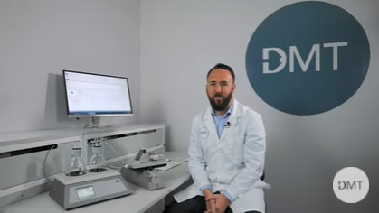 We have completed the video sessions for our Pressure Myograph System - 114P. Our Scientific Product Specialist, Aaron Stupica, gives an in depth look of the DMT Pressure Myograph System – an ideal system for assessment of vascular structure and function of perfused arteries.
  Dr. Pooneh Bagher is the Assistant Professor at theTexas A&M Health Science Center, working on several projects for the National Aeronautics and Space Administration (NASA). Dr. Bagher's lab has studied a range of unique vessels that many researchers have not examined before. Her lab is interested in how blood vessels usually function and how they function differently in either disease states or extreme physiological conditions, including spaceflight.
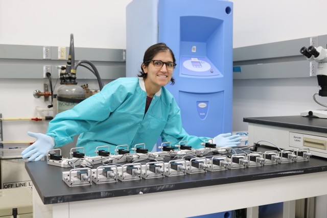
In the past three years, Dr. Bagher and her team have had two distinct areas of focus related to the effects of spaceflight on vasculature. Her group has examined the coronary artery and the angular vein. The latter may play a role in a phenomenon called spaceflight-associated neuro-ocular syndrome, otherwise known as SANS. SANS is a condition that occurs in astronauts when they are in orbit for long-duration missions. Over this period, astronauts start to have changes in their visual acuity. There have been some studies in astronauts on this phenomenon. However, Dr. Bagher and her colleagues first looked at the vascular function in an animal model following space flight. "We were the first researcher sever to study isolated coronary vessels and angular veins following spaceflight," said Dr. Bagher, lead investigator on this part of the project. For Dr. Bagher's most recent work, the animal models spent 38.5 days at the International Space Station before they were brought back to Earth to be studied as part of NASA's Vision Impairment and IntracranialPressure program. Each team member was devoted to looking at a different aspect of the study that was broken down into groups; arteries, lymphatics, and veins. The team collectively looked at the vessels that feed and drain the eyes to see what happened during the space flight. Dr. Bagher's lab has been predominantly using DMT wire myographs in her NASA research. They allow numerous systems to be run in parallel – keeping up with the high demand of samples quickly, which is vital to avoid re-acclimatization of the animal models to Earth's gravity. "We have been using DMT wire myographs since 2015," said Dr. Bagher. When the first project was launched for NASA, Dr.Bagher's Lab held an extreme throughput as they have 28 myograph chambers at their disposal– ranging from single, dual to 4-channel myograph systems. This is essential for collecting many data quickly, which is a must when doing extensive experiments for NASA. The types of experiments Dr. Bagher and her team perform can be very strenuous. The animal models she works with have been taken to space, and the experiments need to be conducted very quickly upon their return. Her entire setup must be fully functional. "We must have equipment that we can coun ton... the DMT wire myograph system provides that consistency," said Dr. Bagher. *Mention of a university affiliation does not constitute an endorsement by Texas A&M University of the content, viewpoint, accuracy, opinions, policies, products, services, or accessibility of any external vendor. Recent Research Comparison of Adrenergic and Purinergic Receptor Contributions to Vasomotor Responses in Mesenteric Arteries of C57BL/6J Mice and Wistar Rats A Mittal, PD Park, R Mitchell, H Fang, P Bagher / Journal of Vascular Research 58 (1), 1-15 Microvascular dysfunction and kidney disease: Challenges and opportunities? S Krishnan, AD Suarez‐Martinez, P Bagher, A Gonzalez, R Liu, ... / Microcirculation, e12661 Endothelial calreticulin deletion impairs endothelial function in aged mice LA Biwer, HR Askew-Page, K Hong, J Milstein, SR Johnstone, E Macal, ... / American Journal of Physiology-Heart and Circulatory Physiology 318 By David Plante | DMT-USA, Inc. | July 2021  A DMT Myograph System is a substantial investment, and maintaining the myograph system is vital to ensure optimal performance, reliability, and output.
Following the maintenance protocols in the user documentation will keep the myograph accurate and reliable for quite a while, with no downtime or failure. However, preventive care reduces the tear and wear of the system, and maintenance can extend the myograph's lifetime. The service check consists of; System dismantles, part cleaning, assembly, lubrication temperature re-compensation of transducers, replacement of internal tubing, calibration, update of firmware *2, functional system test, and documentation. The service is performed by certified technicians only, and the turnaround time is typically 4-5 days. Myograph Maintenance Options
Getting your myograph system serviced by DMT during a valid warranty will add one extra year to the warranty. Contact us for more information *1 annual service for the duration on the agreement *2 when possible  Vascular disease is any abnormalities of blood vessels in the body. According to the American Heart Association data, close to 50% of all adult Americans (~100 million people) have the most common form of the vascular disease known as hypertension. Other forms of vascular disease include atherosclerosis, aneurysms, peripheral artery disease, amongst many others. If left untreated, complications from vascular disease could lead to debilitating illnesses such as heart attack, stroke, and even death in some cases. Although progress is being made in unraveling the mechanisms of vascular disease, more still needs to be done. It is imperative, therefore, that novel vascular disease models are studied in preclinical settings to generate new therapeutics. Myography is a state-of-the-art application in the study of vascular disease ex vivo.
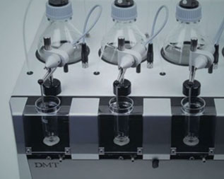
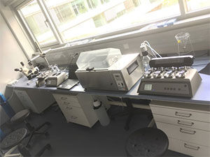
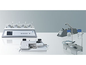
Methodology Myography is simply the study of the velocity of muscle contraction. This technique can be harnessed to measure the force generated when a blood vessel contracts and relaxes under isometric conditions. The generated information can then be used to determine the function and reactivity of blood vessels. Wire myography can be used to study vessels as small as 100 µm to as large as 3 mm in diameter. In brief, vessels are dissected from genetic, transgenic, diseased, or control models, cleaned of adventitia tissues and mounted on wire jaws or steel pins. Mounted vessels are then subjected to a baseline tension, and recordings of force measurement are conducted, and pharmacological effects of drugs can be analyzed. Examples of common vessels used in wire myography studies include conduit arteries such as the aorta, carotid, pulmonary, and some resistance arteries such as mesenteric and cerebral arteries. Pressure Myography is a more physiologically relevant method for assessing functions of small resistance arterioles ex vivo. These small resistance arteries are vital in the modulation of peripheral vascular resistance and blood flow, thereby regulating blood pressure and organ perfusion. To maintain a constant flow, resistance arteries constrict or dilate in response to changes in blood vessel pressure in a process tone as myogenic tone. This autoregulation mechanism results in maintaining a constant flow. To physiologically replicate in vivo vessel conditions in real-time and study resistance arteries using pressure myography, isolated arteries are mounted unto two glass cannulas and pressurized to in vivo pressure. A new development in pressure myography can add a pulse feature to mimic beats per minute (BPM) on the vessel. Under these conditions, vascular and drug effects can then be studied. Using data acquisition software and live tracking, vessel inner and outer diameter and over twenty endpoints are determined under flow or non-flow states. Examples of resistance arteries used in pressure myography include mesenteric, coronary, cerebral arteries with diameters of 100 µm or less. Conclusion What myography technique is best for investigators? Is it wire or pressure myograph, or both? Drawing from my fifteen-year experience as an investigator in hypertension research, I have used both wire and pressure myographs in the same laboratory setting. For large conduit vessels such as the aorta, wire myography is ideal for elucidating the mechanical properties of vessels under isometric conditions. For small resistance arteries, pressure myography is ideal to phenotype near-physiological in vivo conditions. Based on their hypothesis and vessel of interest, it is imperative for the investigator to decide if wire or pressure or both could be incorporated in their scientific tool kit. If you have questions on which methodology is best suited for your study, please contact one of our Scientific Product Specialists. By Dr. Larry Agbor Scientific Product Specialist DMT-USA, Inc. 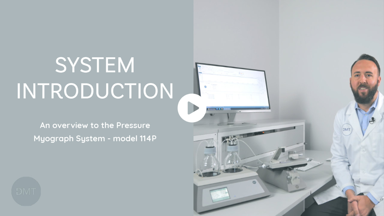 Our Scientific Product Specialist, Aaron Stupica, gives an overview of the DMT Pressure Myograph System – an ideal system for assessment of vascular structure and function of perfused arteries.
Check out the videos on our YouTube Channel, and make sure you subscribe to get the latest releases. 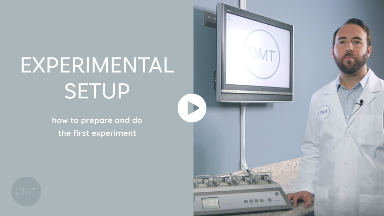 We are excited to share a new video series, which will provide simple video tutorials on using the DMT systems. In the series, we will cover topics from system overview to experimental examples. Our goal is to provide quick videos that complement our written guides and user manuals.
Check out our videos by heading to our YouTube Channel. Make sure you subscribe to get updates when new videos are published. We hope this new video series will be useful in setting up and getting started with your DMT myograph or organ bath system. 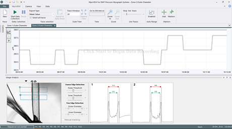 Discover more about our latest features and what was designed to add functionality to the user experience and to make it more intuitive. Live tracking of perfused arteries' vascular reactivity using the DMT Pressure Myograph Systems has never been more simple.
MyoVIEW 5 offers a simple user interface that provides live diameter tracking of perfused arteries. Using accurate edge detection from up to four user-defined zones, MyoVIEW 5 averages the lumen measurements much simpler and more reliable. Wall structure, wall stress, strain, and myogenic tone are measured, and responses to increases in pressure, flow, and drugs are evaluated regarding vascular diameter changes. MyoVIEW 5 provides data acquisition, calculations, and export of data for analysis purposes. It even allows you to create advanced calculations to customize your live traces. Add a script, and traces such as strain is live instantly. Easy and quick use, allowing full control of many parameters and management of data. Display the screen view as you want – there is complete flexibility to arrange windows neat and save your preferred layout.  NEW PARTNERSHIP. We are pleased to announce that we have entered into an agreement with Courteous Pharma CPSB, effective May 1st, 2020. Courteous Pharma CPSB will be our distributor for Malaysia.
Courteous Pharma CPSB has many years of experience, expertise, and knowledge in the fields of biotechnology, medical diagnostic and medical research & development. Partnering with Courteous Pharma CPSB will enhance the local presence of DMT myographs as well as the support of Malaysian myograph users.  Following the outbreak of the Coronavirus and the current pandemic, the safety and security of customers, and employees, and as our top priority.
We have put in place measures to help minimize the risk of Coronavirus to our employees and their families. All office-based employees are working from home, and those in our production that cannot do so are temporarily suspended from work under the government aid schemes. To minimize disruptions, we are continuing to operate to the extent possible and can assist with all matters relating to sales and support. Do not hesitate to get in touch. Stay safe, stay well, and take care. DMT Management 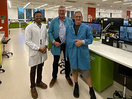 Undergraduate students at the University of Sydney are using live tissue obtained from the local abattoir in their DMT Organ Baths during their pharmacology practical classes.
Senior educators, Dr. Brent McParland and Dr. Nehan Munasinghe have been recognised by the Research Integrity and Ethics Administration at the University of Sydney for their support for the 3Rs (replacement, reduction and refinement). The tissue is obtained from the abattoir where it is kept in appropriate storage for up to 3 months. An additional advantage of the new approach is the significant reduction in lab preparation time and costs. “Using live tissue enables students to view and understand biological variation, improving education outcomes,” said Dr McParland. Nehan (left) and Brent (right) met with DMT CEO Carsten Thorndahl (centre) during his recent visit to Australia. 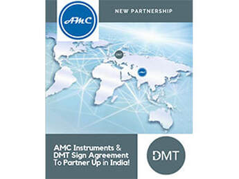
 We are pleased to announce that we have moved our headquarter to a new location! With more space and an optimized workflow, we can serve our customers better and more efficiently. Our new address is:
Danish Myo Technology A/S Rho 14 8382 Hinnerup Denmark
The exhibition begins on Sep. 10 and DMT-USA will be showing several of our myograph systems. Make sure you stop by our booth.
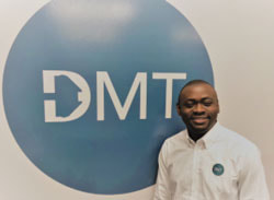
When the Pharmacology team at top-rated Monash University started planning for their new multidisciplinary teaching labs, they looked for alternatives to their traditional, bulky and high-maintenance organ bath systems.
|


|
|

|