|
More about
WIREMyography
|
|

Basic Tissue Properties
|
Pharmacology
|

Physiology Changes & Pathology
|
Vasaoactive Mechanisms
|
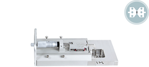
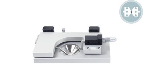
The Wire Myograph allows the examination of small vessels (internal diameter 60 μm - 10 mm) in terms of morphology and responsiveness to hormones and other agonists. The small vessels are mounted as ring preparations by threading them onto two stainless steel or tungsten wires and securing the wires to two supports.
One support is then attached to a micrometer, allowing control of vessel circumference. The other support is attached to a force transducer for the measurement of tension development. The whole preparation is kept in a chamber with physiological salt solution at 37°C, bubbled with oxygen. Vessels maintained in Wire Myographs are viable for several hours.
Following mounting and equilibration, the passive length-tension relationships of the vessels are determined; a normalization procedure. During the actual experiments, vessels' circumference are kept constant and vessels are examined under isometric conditions. Compounds are added directly to the chamber and vessel tension is monitored. Furthermore, it is possible to compare vessels from patients or test groups with those of control, not only in terms of vessel reactivity to various compounds but also in terms of morphology.
The following lists are a few of the established areas of investigation for Wire Myograph systems. Many more investigation possibilities for vascular and other smooth muscle may be added through the imagination of researchers such as yourself.
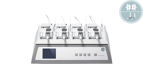
Wire Myograph Systems
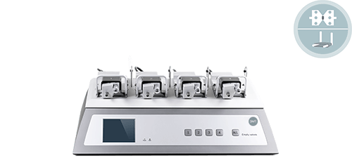
Auto. Multi Wire Myograph System - 630MAMaking it easier… the 4-channel system adds the ease of automating the normalization procedures so that calculations and preload tension is easily set. Following mounting and equilibration, passive length-tension relationships are determined by a standardized procedure.
|