|
More about
PRESSUREMYOGRAPHY
|

|

Basic Tissue Properties
|

Pharmacology & Physiology
|

Pathology
|

Vasaoactive Mechanisms
|
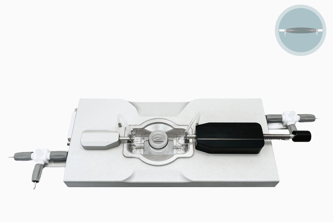
Pressure Myograph Systems are used to measure small arteries, veins, and other vessels' physiological function and properties. The system also allows studying pharmacological effects of drugs and other vasoactive compounds on small isolated vessels under near-physiological conditions. In these systems, vessels retain many of their in vivo characteristics.
In pressure myography, an intact small segment of an artery or vein is mounted onto two small glass cannula and pressurized to a suitable transmural pressure. This near physiological condition permits the investigation of intrinsic (myogenic) responses, which can be extrapolated to the entire vascular bed's in vivo behavior (auto-regulation).
Various pharmacological agents can then be studied by adding these to the superfusate or luminal solution. Both constriction and dilation can be readily measured as changes in the diameter of the preparation via digital video edge-detection. Since intrinsic myogenic constriction is present, the endothelium's role and function for this phenomenon can be studied.
Specialized versions of pressure myograph chambers can accommodate particular research need like imaging (confocal) or electrophysiology.
Overall, the pressure myograph technique is a very powerful tool in the dedicated vascular physiologist or pharmacologist's hands.
The following lists are a few of the established areas of investigation for the pressure myograph systems. In the future, many more areas will be added to these lists through ongoing product development and the research performed by myograph users.
Pressure Myograph Systems
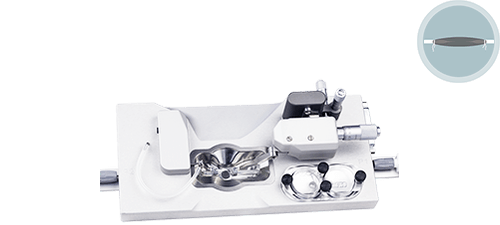
Pressure Pulsation Myograph 112PPIdeal system used to study the structure and function of isolated sections of small vessels (diameter >40μm) under near-physiological conditions. Vessel diameters can be measured in response to pharmacological and physiological stimuli. In addition, this system allows simulation of pulsations between 50 and 600BPM with a pressure difference up to 60mmHg.
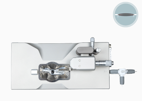
Pressure Myograph 114PBThe Blind Sac Pressure or Perfusion Myograph System - 114PB is a system used to study the structure and function of isolated sections of small vessels (diameter >40 μm) under near physiological conditions. Vessel diameters can be measured in response to pharmacological and physiological stimuli.
Primary area of research is physiological responses under myogenic tone. |
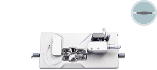
Pressure Myograph 114PUsed to measure the physiological function and properties of small arteries, veins, and other vessels. Study pharmacological effects of drugs and other vasoactive compounds under near-physiological conditions. An intact segment is mounted onto two small glass cannulas and pressurized to a suitable transmural pressure.
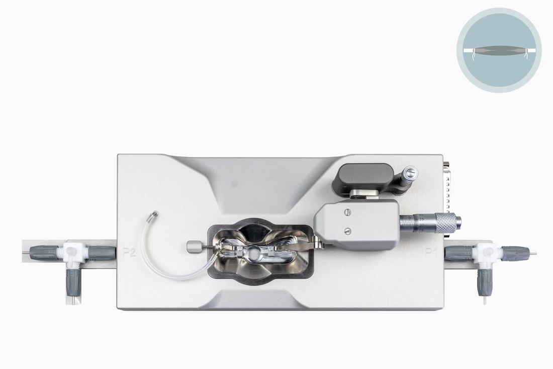
Pressure Myograph 114PNThe Pressure Myograph System - 114PN has less optional features while still allowing experiments on pressurized small arteries, veins, and other vessels. This specific model is for researchers who want to design their pressure and flow studies without measuring the longitudinal force. A small, intact segment of an artery or vein is mounted onto two small glass cannulas and pressurized to a suitable transmural pressure. The near-physiological conditions permit the investigation of myogenic responses and flow-mediated dilation in response to pharmacological agents and other vasoactive compounds.
|
Pressure Myograph 110PXLThe Pressure Myograph System - 110PXL is used to measure large arteries, veins, and other vessels physiological function and properties. The 110PXL chamber, in combination with low-power objectives, can be used for the study of large artery reactivity and/or compliance by measuring diameter changes under physiological pressures, which are precisely controlled through the system interface.
|
