 🚀 Exciting News! We're thrilled to announce the relaunch of our 560TP Tissue Puller System!
The 560TP is a user-friendly tensometer designed for quick and easy determination of tensile strength in tissues. With the MyoPULL software, users can swiftly evaluate characteristics such as compliance, stress-strain, and fatigue through real-time plotting of the length-tension relationship of the sample tissue. This advanced system streamlines your research process, providing accurate and efficient results. Get ready to elevate your tissue analysis to the next level! 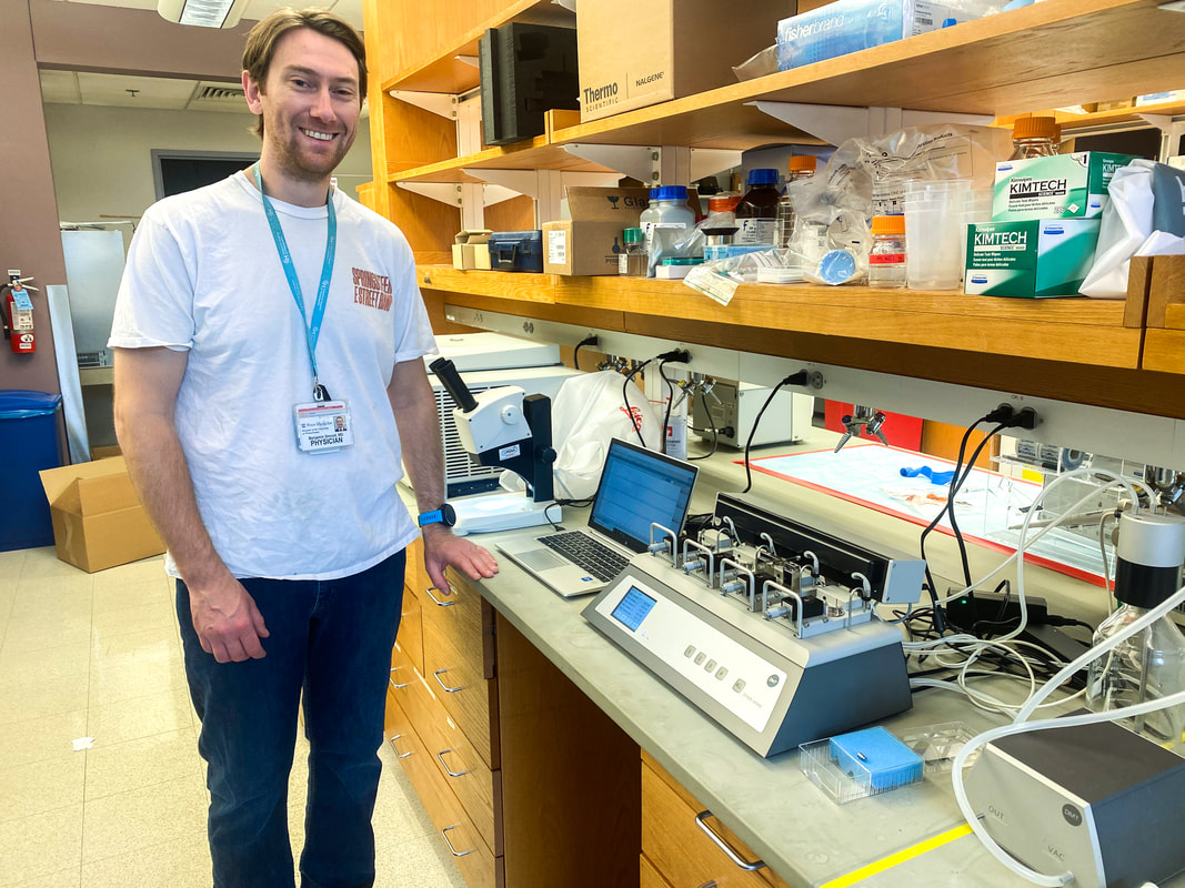 We had a wonderful visit with Benjamin Smood, MD at Penn Medicine, University of Pennsylvania Health System! Excited to share the successful installation of the 620M Wire Myograph paired with our Buffer Filler System. This state-of-the-art equipment will empower researchers and students to delve deeper into vascular physiology, advancing our understanding of cardiovascular health and disease. Kudos to our Sr Scientific Product Specialist, Larry Agbor, Ph.D, M.S for aiding in the installation!
#WireMyography #Research #CardiovascularScience #PennStateUniv 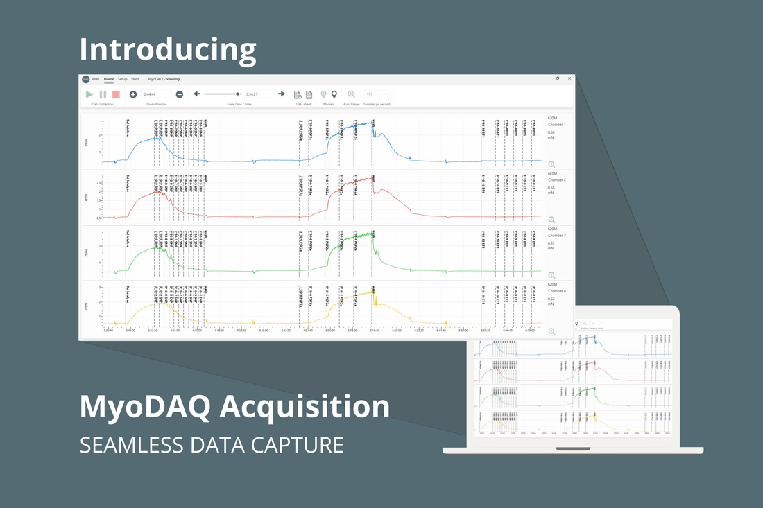 We are pleased to announce the launch of our new proprietary data acquisition software, MyoDAQ! MyoDAQ offers a simplified data acquisition application intended for use with DMT wire myographs and organ bath systems. This software provides a streamlined, user friendly approach for data collection and analysis.
By highlighting only necessary features, researchers experience improved ease-of-use, better navigation and easier control for data analysis. To learn more about this software, visit https://www.dmt.dk/myodaq.html or contact a DMT representative! #dataanalysis #dataacquisition #wiremyography #organbath 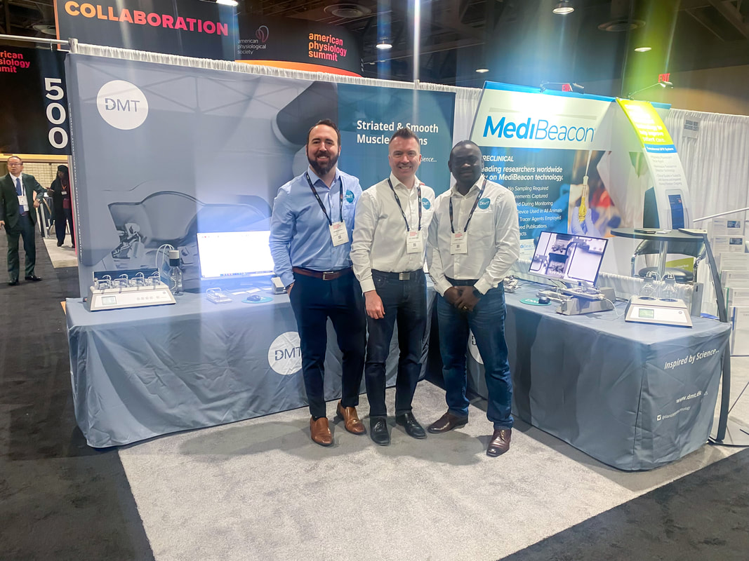 Having a great first day at The American Physiology Summit @APSPhysiology. We love seeing a lot of familiar faces and connecting with everyone. Make sure to stop by our booth (#520) and say hello. We look forward to seeing you! #APS2024 #WeArePhysiology
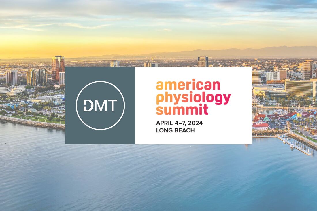 Just one week until the American Physiology Summit! We look forward to attending and connecting with you all there. Stop by our booth (#520) for the latest updates on our myograph systems and get a sneak peek of our new and simplified data acquisition software, developed explicitly for organ bath and myograph systems. See you soon!
#APS2024 In a few weeks, at the American Physiology Summit, stop by our booth (#520) and get a sneak peek of our new and simplified data acquisition software, developed explicitly for organ bath and myograph systems. 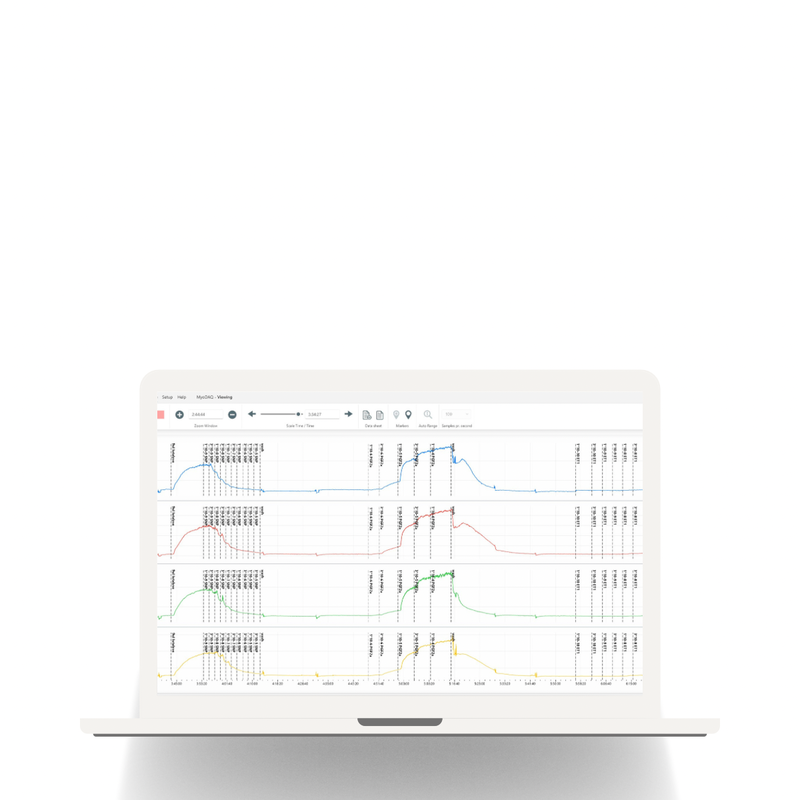 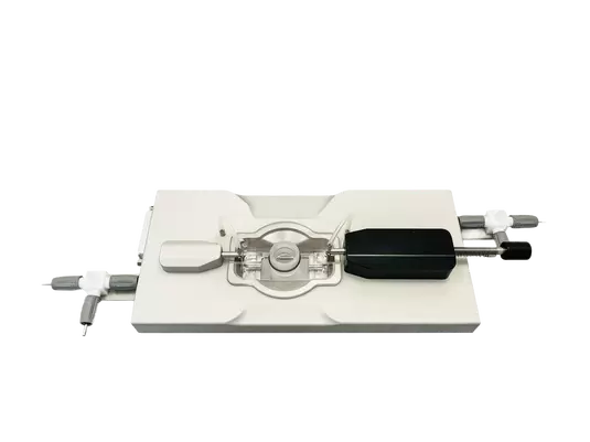 The Pressure Myograph System - 110PXL is used to measure large arteries, veins, and other vessels physiological function and properties. The 110PXL chamber, in combination with low-power objectives, can be used for the study of large artery reactivity and/or compliance by measuring diameter changes under physiological pressures, which are precisely controlled through the system interface.
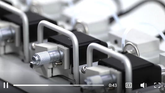
The system comes with mounts of 1.5mm, 3mm, and 4mm for mounting vessels between 1.5-6.0mm. There is no force transducer on the standard chamber, but it can be added. This system is a great option for expanding pressure myograph capabilities! 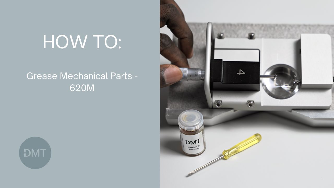 In this video, you will be learning about how to grease important mechanical parts on the Multi-Myograph System 620M.
Product Link: Multi-Myograph System 620M To learn more visit our Resources Page For more information visit us at www.dmt.dk or email [email protected] to speak to a representative. 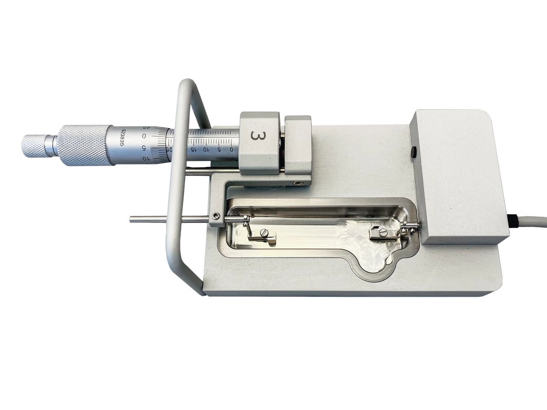 Introducing the 820XL chamber! This robust organ bath chamber offers myograph users a larger tissue capability while still maintaining the compact features of the organ bath interface.
Users will be able to work with rings larger than 500µm and strips up to 45mm in length, giving an ideal range for those experimenting with larger vessels. For more information on the 820XL Chamber, visit: https://www.dmt.dk/organ-bath-820mo.html 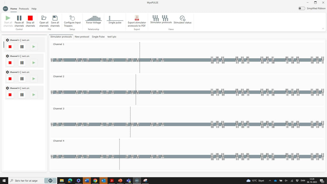 We’re thrilled to release an updated version of MyoPULSE software for our current simulators. Packed with powerful features, enhanced performance and a new improved user experience, this release takes a big step forward.
Here’s some of the new features…
Interested in upgrading your current version of MyoPULSE - contact us  APAC Scientific is showcasing DMT's latest Myograph and Organ Bath systems at the ASCEPT Annual Scientific Meeting, 20-23 November, 2023 in Sydney, Australia.
 DMT will be represented by China Gate Scientific (CGS) at the upcoming World Congress of Microcirculation, September 20-24, in Beijing, China.
Cutting-edge, innovative myograph systems will be on display, and the CGS team will be on hand to provide insights, answer questions and offer recommendations to enhance your vascular reactivity studies. 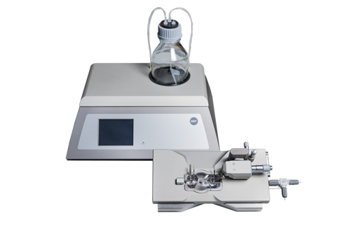 We are happy to announce the NEW 114PB Pressure Myograph System! This cost effective system is able to provide consistent and reliable results for pressurized vessel studies only. Our Scientific Product Specialist, Aaron Stupica gives an in depth look of the simplified pressure system which can be found in the training video series!
 DMT is excited to exhibit at the upcoming ESM meeting in Aarhus, Denmark. We'll be displaying the latest innovative systems and recently added pressure myograph chambers. Make sure to stop by our booth.
 We are getting ready to attend the American Physiology Summit being held in Long Beach, CA April 20-24. Stop by and learn more about our new pressure myograph chambers.
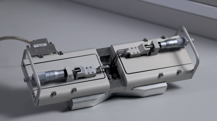
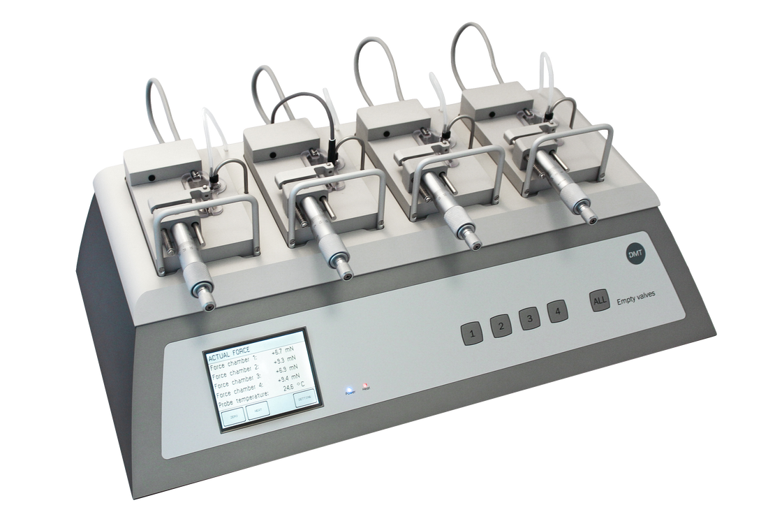
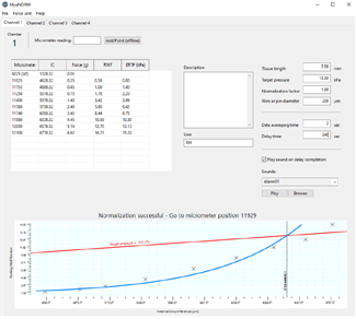
 Our Scientific Product Specialist, Aaron Stupica gives an in depth look of the DMT Automated Wire Myograph System. This 4-channel system allows the study of any tubular segment's vascular functionality while adding the feature of automated normalization procedures so calculations and preload tension are easily set. Click the links in the description to watch our full video training guides and get more in depth information on our myograph systems!
 DMT is excited to exhibit at the European Muscle Conference in Prague, Czech Republic. You will find our Scientific Product Specialist, Dr. René Hummel at our booth – ready to show you the variety of our systems for muscle physiology research.
 Announcing the DMT "How To" video series!
Ever wondered how you mount an artery on a DMT myograph system? Or how mounting supports are changed? Well...we have just released a series of "how to" videos that will teach you a variety of techniques and reference the use of DMT myograph systems. Make sure to subscribe to our YouTube page so you don't miss out on any new videos and content that we release! #DMT #Myograph 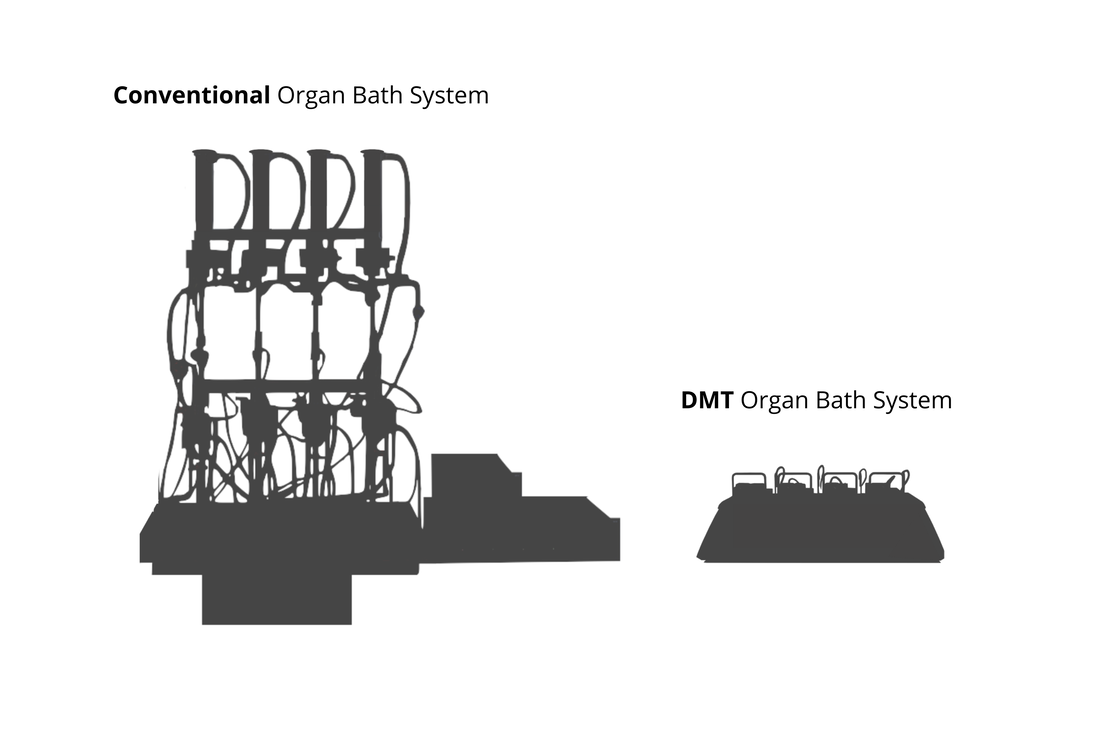 Yes, it does...when it comes to efficiency and output in a research laboratory.
Gone are the days when the organ bath system took up an entire bench top. The DMT Organ Bath system is a compact and robust design, where the footprint is so much smaller that you can fit several systems within the footprint of the conventional organ bath. This allows you to save a tremendous amount of valuable lab space and maximize your throughput. Minimize your equipment footprint while maximizing results. For more information on the DMT Organ Bath System, check out our 820MO page! #DMT #DanishMyoTechnology #BasicResearch #OrganBath #IsolatedTissue 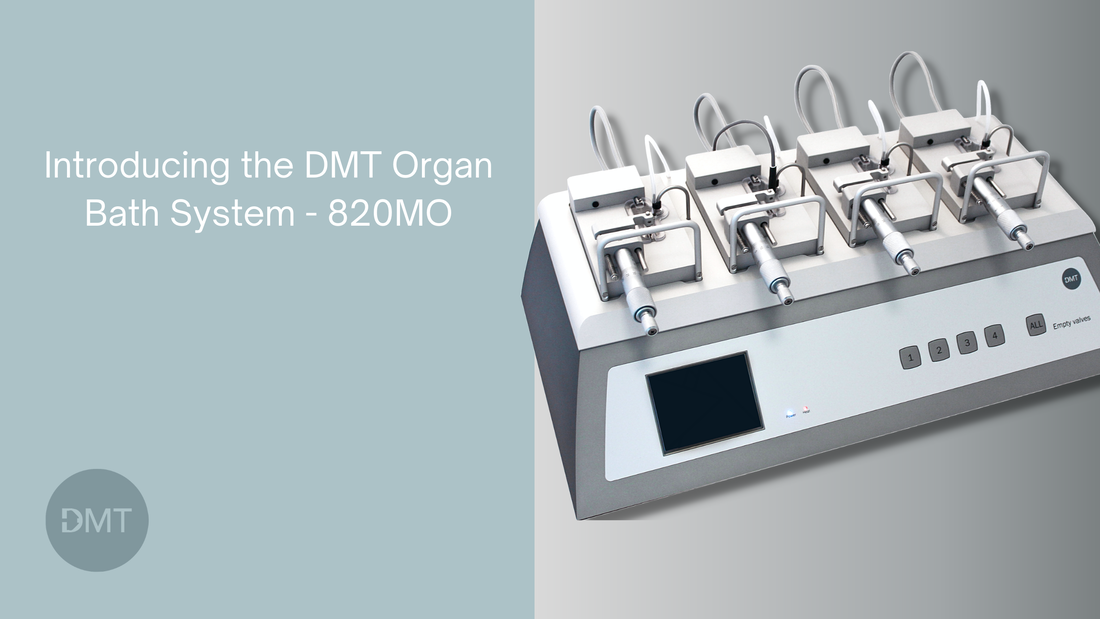 Organ baths are a well-known, classic experimental setup commonly used for reactivity studies of ex vivo tissue preparations. With the introduction of the DMT Organ Bath – 820MO, we combine decades of knowledge from myography and conventional organ bath setups into a modern, state-of-the-art system that enhances productivity. The robust yet compact design adds value to users with built-in features such as oxygenation, buffer drainage, calibration procedures, and heating. It even has selectable force ranges for the type of tissue used.
 Interesting publication in which the DMT MyoDYNAMICS Muscle Strip System - 840MD were used. Read the full article at MDPI/biomedicines
  We have RELAUNCHED the 420A Dual Chamber Wire Myograph System! This was one on the earliest Myographs released by DMT in the early 1980's and it has been updated and reintroduced. This is a perfect system for side by side vessel comparison studies, small working volume and reproducible and reliable experiments. 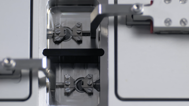 The durability of the build and materials allow for heavy use and reproducible experiments. This system has been built for optimal performance which will last many years.
For more information about the 420A Dual Chamber Wire Myograph System, visit the 420A product page.
|


|
|

|