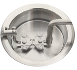
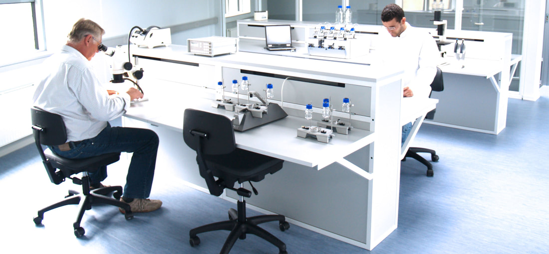
 Vascular disease is any abnormalities of blood vessels in the body. According to the American Heart Association data, close to 50% of all adult Americans (~100 million people) have the most common form of the vascular disease known as hypertension. Other forms of vascular disease include atherosclerosis, aneurysms, peripheral artery disease, amongst many others. If left untreated, complications from vascular disease could lead to debilitating illnesses such as heart attack, stroke, and even death in some cases. Although progress is being made in unraveling the mechanisms of vascular disease, more still needs to be done. It is imperative, therefore, that novel vascular disease models are studied in preclinical settings to generate new therapeutics. Myography is a state-of-the-art application in the study of vascular disease ex vivo. Methodology Myography is simply the study of the velocity of muscle contraction. This technique can be harnessed to measure the force generated when a blood vessel contracts and relaxes under isometric conditions. The generated information can then be used to determine the function and reactivity of blood vessels. Wire myography can be used to study vessels as small as 100 µm to as large as 3 mm in diameter. In brief, vessels are dissected from genetic, transgenic, diseased, or control models, cleaned of adventitia tissues and mounted on wire jaws or steel pins. Mounted vessels are then subjected to a baseline tension, and recordings of force measurement are conducted, and pharmacological effects of drugs can be analyzed. Examples of common vessels used in wire myography studies include conduit arteries such as the aorta, carotid, pulmonary, and some resistance arteries such as mesenteric and cerebral arteries. Pressure Myography is a more physiologically relevant method for assessing functions of small resistance arterioles ex vivo. These small resistance arteries are vital in the modulation of peripheral vascular resistance and blood flow, thereby regulating blood pressure and organ perfusion. To maintain a constant flow, resistance arteries constrict or dilate in response to changes in blood vessel pressure in a process tone as myogenic tone. This autoregulation mechanism results in maintaining a constant flow. To physiologically replicate in vivo vessel conditions in real-time and study resistance arteries using pressure myography, isolated arteries are mounted unto two glass cannulas and pressurized to in vivo pressure. A new development in pressure myography can add a pulse feature to mimic beats per minute (BPM) on the vessel. Under these conditions, vascular and drug effects can then be studied. Using data acquisition software and live tracking, vessel inner and outer diameter and over twenty endpoints are determined under flow or non-flow states. Examples of resistance arteries used in pressure myography include mesenteric, coronary, cerebral arteries with diameters of 100 µm or less. Conclusion What myography technique is best for investigators? Is it wire or pressure myograph, or both? Drawing from my fifteen-year experience as an investigator in hypertension research, I have used both wire and pressure myographs in the same laboratory setting. For large conduit vessels such as the aorta, wire myography is ideal for elucidating the mechanical properties of vessels under isometric conditions. For small resistance arteries, pressure myography is ideal to phenotype near-physiological in vivo conditions. Based on their hypothesis and vessel of interest, it is imperative for the investigator to decide if wire or pressure or both could be incorporated in their scientific tool kit. If you have questions on which methodology is best suited for your study, please contact one of our Scientific Product Specialists. By Dr. Larry Agbor Scientific Product Specialist DMT-USA, Inc. Comments are closed.
|
�
|


